i BT12


i BT12
The Vivid i is based on GE’s TruScan Architecture, which is common to all GE Ultrasound systems

System Architecture
The Vivid i is based on GE’s TruScan Architecture, which is common to all GE Ultrasound systems, EchoPAC*PC Workstation, software and network solutions
User Interface – Operator Keyboard
Easy-to-learn user interface with intelligent keyboard, Keyboard with application specific push buttons for primary controls, Interactive back-lighting of application-specific push buttons
Dimension
Height: 64 mm (2.5 in), Width: 358 mm (14.2 in), Weight: approximately 5.8kg (12.7 lbs)
Display Screen
High-resolution, flat 15-inch TFT LCD screen, Display size: 1024 x 768 pixels with 260,000 simultaneous colors available, Scanner software supports display resolution of 800 x 600 pixel.
Data Acquisition
Application-Specific Channel Architecture: the Vividi employs a flexible digital beam-former architecture capable of using up to 99,740 effective channels depending on specific application requirements.
Color Doppler Imaging
Digital signal processing power maintains high frame rates with large ROIs even for very low PRF settings, Variable Region of Interest (ROI) size in width and depth
Imaging Solutions
The Vivid i is designed for cardiovascular imaging, abdominal, small parts, perioperative monitoring and 2D ICE imaging
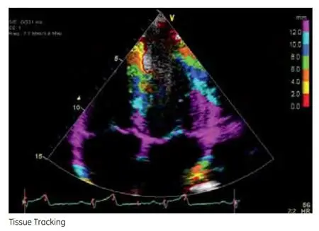
Clinical Image 1
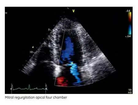
Clinical Image 2
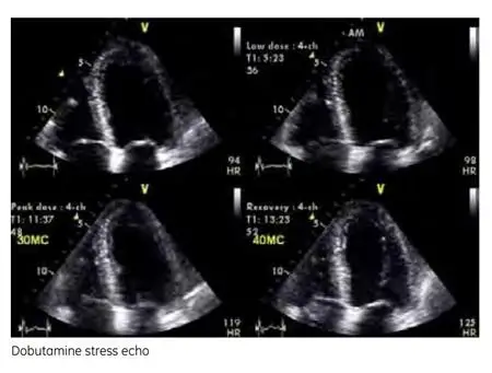
Clinical Image 2

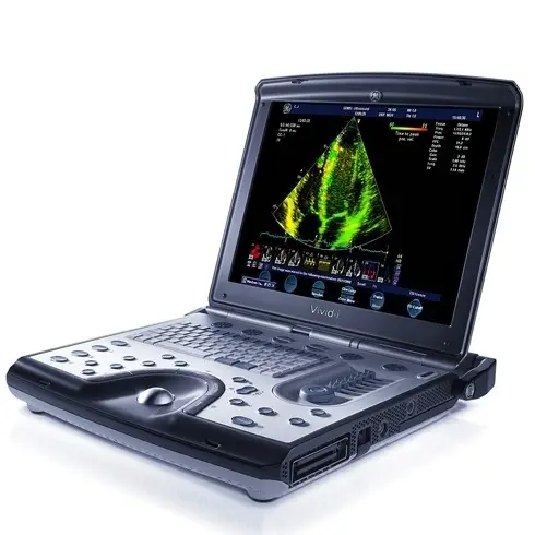
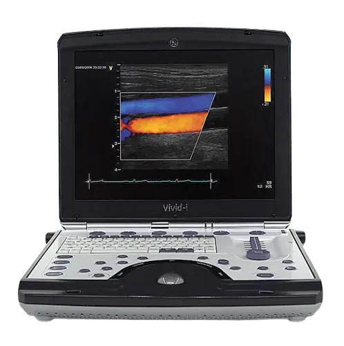
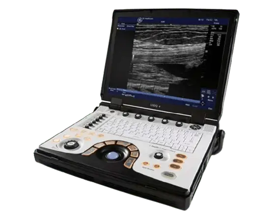
About GE Vivid i BT12
The Vivid i is a high-performance, battery-operated, ultra-portable diagnostic Ultrasound systems providing exceptional image quality. The Vivid i is designed for cardiovascular imaging, abdominal, small-parts, perioperative monitoring and 2D ICE imaging. The Vivid i is based on GE’s TruScan Architecture, which is common to all GE Ultrasound systems, EchoPAC*PC Workstation, software and network solutions. It features a software-driven PC-based platform, raw data storage with advanced post-processing capabilities, seamless DICOM** standard connectivity and compatibility with the GE family of Cardiovascular Ultrasound Systems. Innovative tools offer advanced connectivity, remote monitoring and consultation for improved productivity and quality of care.
Compatible Probes
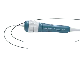
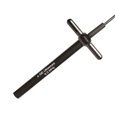
AcuNav 10F
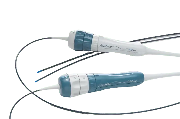
Bandwidth: 4.5 – 11.5 MHz
Footprint: 90 cm length
Field of View: 49.6 mm
GE 3Sc-RS
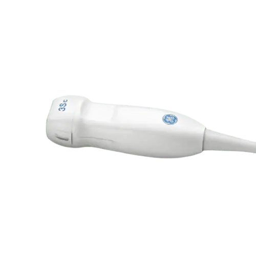
Bandwidth: 1.3 – 4.0 MHz
Footprint: 18 x 24 mm Footprint
Field of View: 90° Field of View
GE 8L-RS
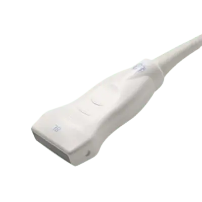
Bandwidth: 4 – 13 MHz
Footprint: 14 x 48 mm
Biopsy Guide: Multi angle
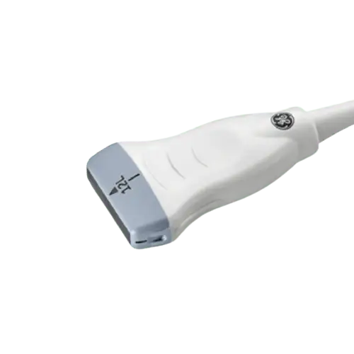
GE i12L-RS

Bandwidth: 1 – 5 MHz
Field of view: 90o
Number of elements: 3040
GE 6Tc-RS
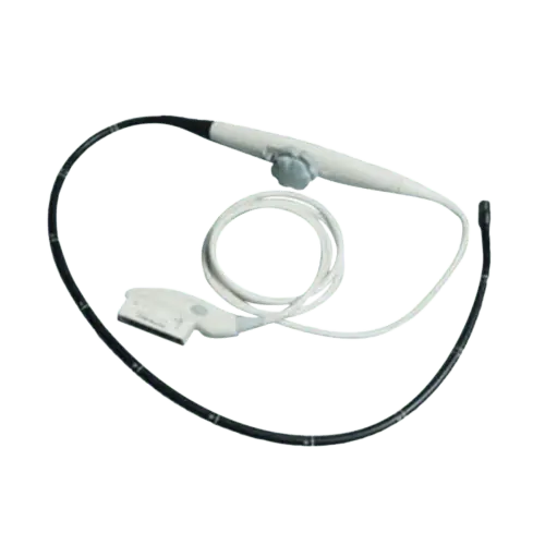
Bandwidth: 2.9-8.0 MHz
Footprint: length 45 mm
Depth of field: 20cm
GE 7S-RS
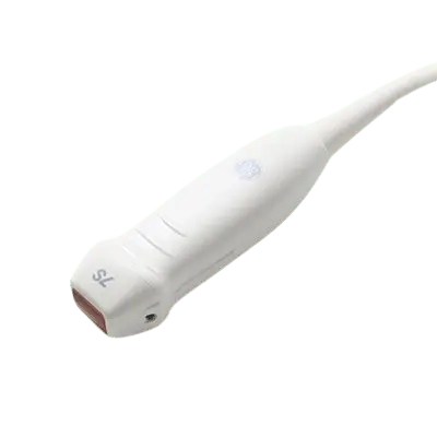
Bandwidth: 3.5 – 8 MHz
Footprint: 15 x 21 mm
Applications: Pediatric Cardiac
GE 10S-RS
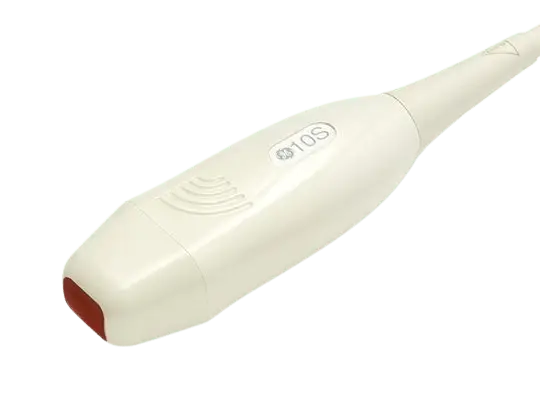
Bandwidth: 5 – 11.5 MHz
Footprint: 10 x 14 mm (Vivid i, q)
Applications: Neonatal Cardiac
AcuNav 8F
Bandwidth: 3 – 10 MHz
Footprint: 7 mm
Applications: Intracardiac Echo
GE 9L-RS
Bandwidth: 3.33 – 10 MHz
Footprint: 14.1 x 53 mm
Field of View: 49.6 mm
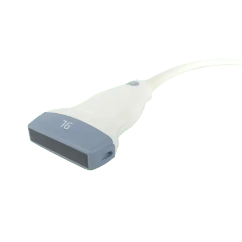
GE 12L-RS
Bandwidth: 6 – 13 MHz
Footprint: 14 x 48 mm Footprint
Biopsy Guide: Available
GE 4C-RS
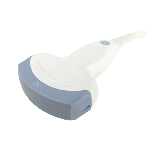
Bandwidth: 1.8 – 6 MHz
Footprint: 17 x 65 mm Footprint
Field of view: 90o
GE 8C-RS
Bandwidth: 4.7 – 11 MHz
Footprint: 26 x 10 mm Footprint
Imaging Modes: 2D, harmonics
GE e8C-RS
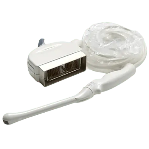
Bandwidth: 4 – 10 MHz
Footprint: 26 x 10 mm
Field of View: 49.6 mm
GE 9T-RS
Bandwidth: 4 – 10 MHz
Footprint: 10.7 x 7.5 mm
Applications: Pediatric Cardiac
GE 3S-RS
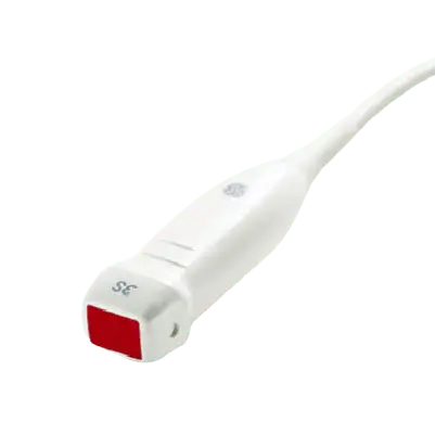
Bandwidth: 1.5 – 3.6 MHz
Footprint: 18 x 24 mm
Biopsy Guide: Multi angle
GE 6S-RS
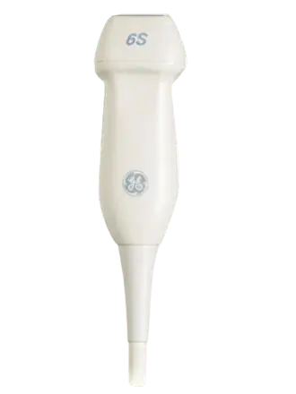
Bandwidth: 2.7 – 8 MHz
Footprint: 17 x 23 mm
Field of view: 90-degree
GE 12S-RS
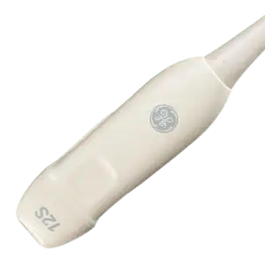
Bandwidth: 5.0 – 11MHz
Footprint: 13 x 18mm
Field of Depth : 12 cm
Downloads


Have a Question?
sales@theultrasoundsource.com support@theultrasoundsource.com
Call US
(888) 514-0911

Ready to Buy?

