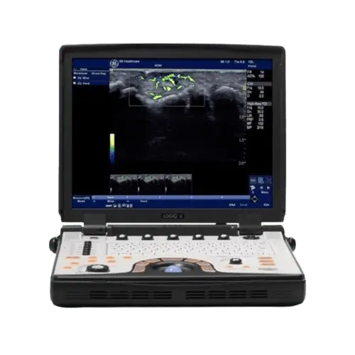
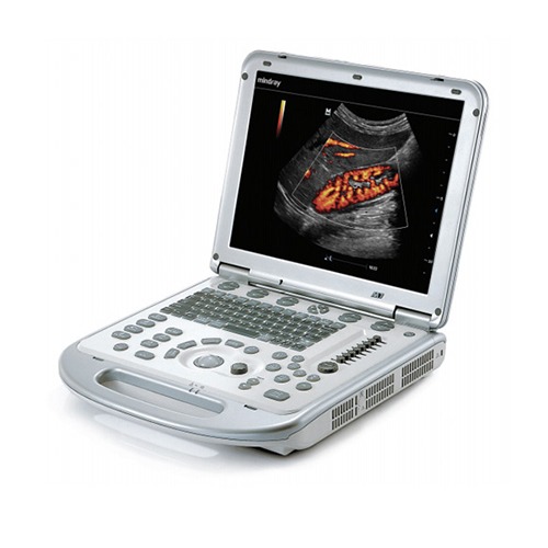
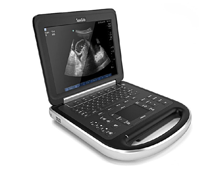
e BT12


The GE Logiq e BT12’s high-resolution image quality gives you the information you need to help make quick decisions with confidence

Point of Care versatility
Amazingly Accurate Anatomy: Our Tissue Differentiation comes from the combination of our proprietary imaging technology, beamformer and ultra-high frequency L8-18i-RS transducer.
Operator Keyboard
Alphanumeric Keyboard, Ergonomic Hard Key Operations, Integrated Recording Keys for, Peripheral and DICOM Devices
Dimension
Height: 70mm(2.75in) console only, Width: 340mm(13.38in), Depth: 346mm(11.63in) console only
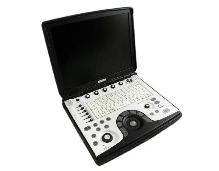
15-inch TFT LCD screen
High Resolution 15 inch Color LCD, Over 1,000 frames or over 60 seconds CINE Memory (64 MB) depend on FOV, Scanning Lines etc.
eSmart Trainer
eSmart Trainer provides modules showing basic scanning techniques, with graphics of probe position, anatomy and example clinical image
Standard Features
160 GB Hard Drive, External DVD R/W storage, Loop storage—from live scanning and from memory, Automatic Optimization
Imaging Solutions
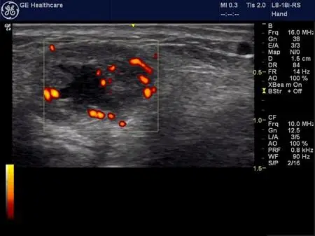
Power Doppler Imaging (PDI) sensitivity detects slow blood flow in both small and large vessels. B-Steer + Needle Recognition delivers accurate detail needle, anatomy and motion even in Colorant Power Doppler.
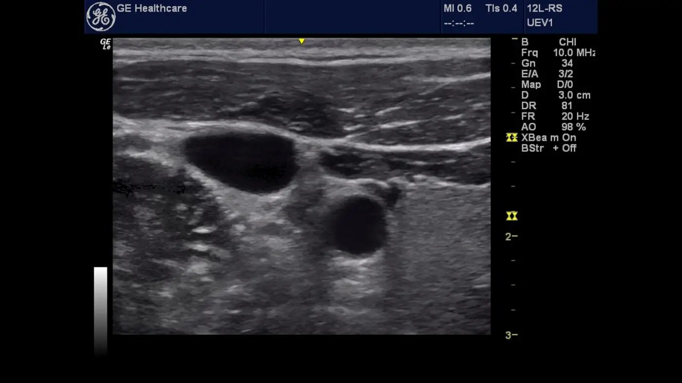
Clinical Image 1
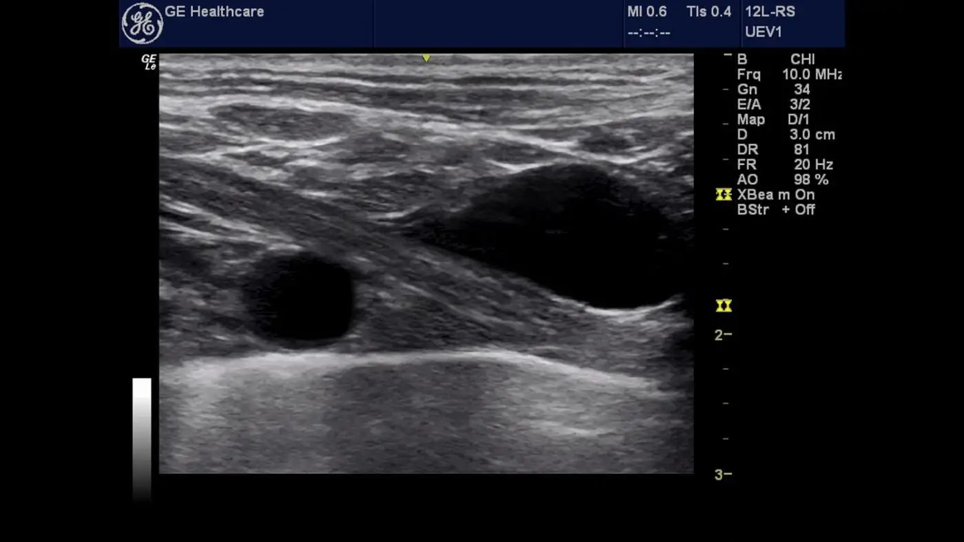
Clinical Image 2
Clinical Image 3

About GE Logiq e BT12
GE LOGIQ e BT12 gives physicians the power to expand from routine to advanced ultrasound imaging. GE Healthcare’s leadership compact system is designed for general imaging, musculoskeletal, anesthesiology, interventional, emergency, and critical care applications. A variety of premium technologies help care for a broad spectrum of patients, from superficial to dynamic or deep imaging at the point of care. Our Tissue Differentiation comes from the combination of our proprietary imaging technology, beam former and ultra-high frequency L8-18i-RS transducer. This helps you detect subtle changes in anatomy, minimal amounts of fluid and small structures.
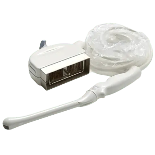
Compatible Probes
GE 4C-RS
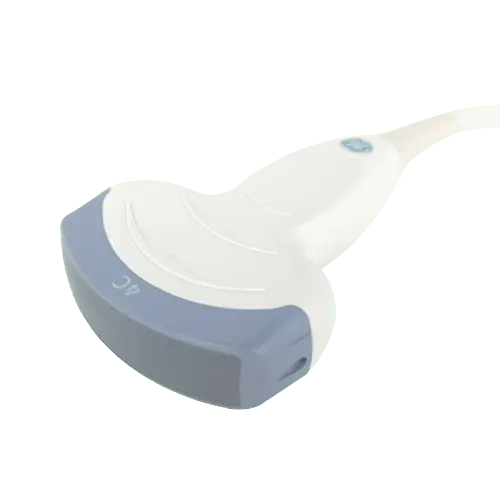
Bandwidth: 2 – 5.5 MHz
Footprint: 65 x 16 mm
Field of view: 90o
GE 8C-RS
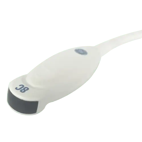
Bandwidth: 4 – 11 MHz
Footprint: 60.7 x 16 mm
Footprint: 26 x 10 mm
GE 6S-RS
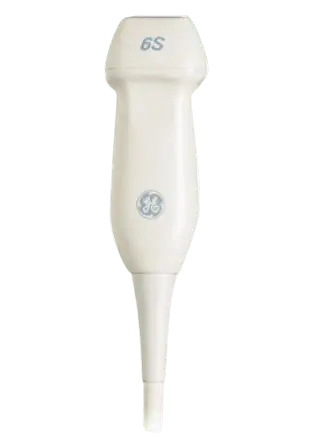
Bandwidth: 2.5 – 8 MHz
Footprint: 16.8 x 23.5 mm
Field of view: 90-degree
GE E8C-RS
Bandwidth: 4 – 11 MHz
Applications: Obstetrics, Gynecology
Field of View: 49.6 mm
GE 3S-RS
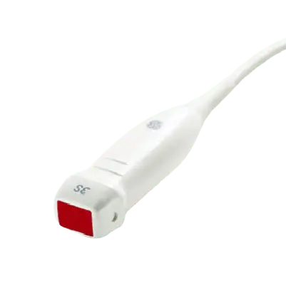
Bandwidth: 1.5 – 4 MHz
Footprint: 21 x 15 mm
Biopsy Guide: Multi angle
GE L8-18i-RS
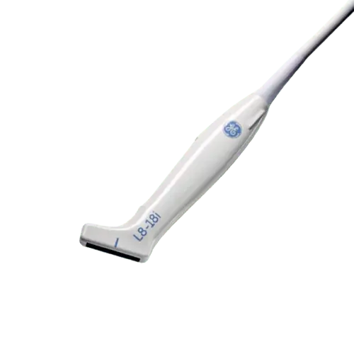
Bandwidth: 6.7–18.0 MHz
Footprint: 11.1 x 34.8 mm
Field of View: 49.6 mm
GE 8L-RS
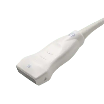
Bandwidth: 4 – 13 MHz
Footprint: 48 x 8mm
Transducer Type: Linear
GE 12L-RS
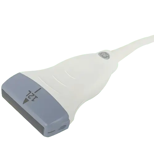
Bandwidth: 5 – 13 MHz
Imaging Modes: 2D, SRI, Harmonics
Footprint: 48 x 7 mm
GE 6Tc-RS
Bandwidth: 2.9-8.0 MHz
Footprint: L45 x W14 (x H12.5) mm
Depth of field: 20cm
GE i12L-RS

Bandwidth: 1 – 5 MHz
Field of view: 90 degree
Number of elements: 3040
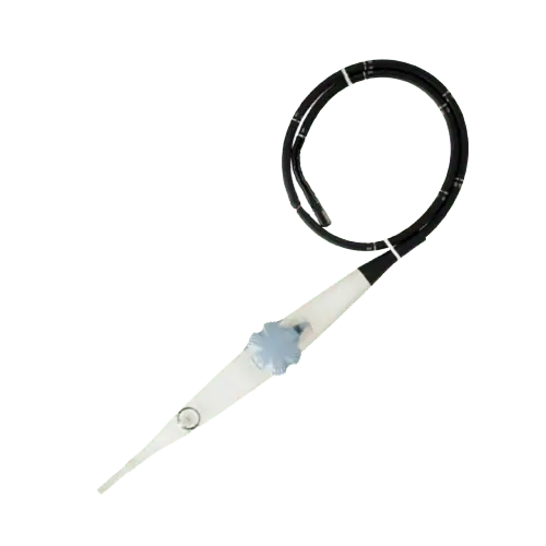
GE i/T739-RS
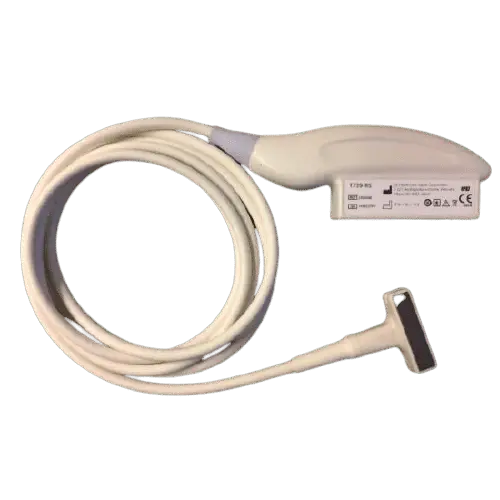
Bandwidth: 3.5 – 9.5 Mhz
Footprint: 44 x 10 mm
Biopsy Guide: Multi angle, Reusable
GE P2D Big Connector
Bandwidth: 2.0 MHz
Footprint: 17mm diameter
Applications: Cardiac, Vascular

Related Ultrasound Machines


Have a Question?
sales@theultrasoundsource.com support@theultrasoundsource.com
Call US
(888) 514-0911

Ready to Buy?

