Steatotic liver disease is also known as fatty liver disease. This is when fat builds up in the liver. The problem can get worse as time goes by. You may not feel sick at first or see any signs of fatty liver disease. Even if you feel well, the liver can change in ways you do not feel. These changes can lead to cirrhosis.
Male rats are often used by scientists to look at steatotic liver disease. They are a good model for fatty liver study because they show clear patterns of liver fat build-up and how their bodies react to changes. This helps researchers use 3D ultrasound to watch how the disease gets worse, see different imaging features, and try out new treatments.
One big thing about steatotic liver disease is that fat can build up in the liver. But having a fatty liver is more than just where the fat sits. Changes in how the body and other things work together also play a part in this chronic liver disease. A lot of things can come together to give someone fatty liver disease or steatotic liver disease.
3D multiparametric ultrasound is good for studying steatotic liver disease in rats. But there are some limits to using this test. For example, it might not always catch small cell changes or early liver damage besides the fat build up. The skill of the person doing the test and the type of ultrasound can also change how clear the results are. Things like how big the rat is and how well the ultrasound machine works can also change the accuracy of what people see.
Doctors use a multiparametric ultrasound when they want to check for fatty liver disease. This ultrasound lets them see the liver in more detail. A multiparametric ultrasound can help find fatty liver and show what is going on. While a liver biopsy is often used, it gives more answers about liver disease and can help you understand it better.The main issue here is to notice these changes in a quick and correct way. A team at POSTECH (Pohang University of Science and Technology) used multiparametric ultrasound to show how blood flows in the liver. This method can help people find liver disease and steatotic liver disease in a new way. With this, you can spot these problems earlier and see them more clearly. The latest studies say that 3D ultrasound is very good at finding liver fat build-up in rats. This technology lets you measure changes without cutting into the body and clearly see fatty changes. All this makes it a good tool for early diagnosis in the lab when checking for liver disease.
Understanding disease progression and detection
Steatotic liver disease (SLD) is the most common type of chronic liver disease in the world. This starts when there is too much fat in the liver. Over time, this can make the liver swell. Liver fibrosis can also develop if this is not stopped early. When you do not get care for SLD, your liver could turn to cirrhosis. In the worst cases, this disease can even turn into liver cancer. This is why it is very important to find SLD early, get checked, and take care of your liver disease.
Many doctors use regular ultrasound. It is a fast way to see fat in the liver. However, ultrasound can have some problems. The test is not always right. The results can also change if someone else does the scan. Magnetic resonance methods, like MRI, often give better results than ultrasound.
Innovative ultrasound technology
To solve these problems, the POSTECH team, led by Professors Chulhong Kim and Yong-Joo Ahn, spent time looking at small microvascular changes in the liver. These changes take place as steatotic liver disease gets worse. The group talked about what they found out about liver disease in a paper that was put out in Nature Communications.
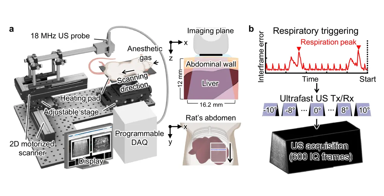
The team made an ultrasound system. This system lets people see blood vessels in the liver as a 3D picture. It has a significant role because it works like seeing city traffic from above. With this, you can spot changes in the blood vessels right away. For example, you will know if a vessel is blocked or if there is any change in the shape of the vessels.
At the center of this is ultrafast Doppler imaging. This type of ultrasound can take thousands of pictures each second. It helps you see blood as it moves in very small blood vessels. Some of these blood vessels are even thinner than a hair.
The team used this and some other ultrasound tools. These other tools help them see fat in the liver and also check the shape of tissues. A few of these, like attenuation imaging (ATI) and acoustic structure quantification (ASQ), are important in research.
Together, these parts help make a three-dimensional, multiparametric ultrasound system. This ultrasound system can show blood flow and tissue at the same time. So, it is a good tool for clinicians. It uses multiparametric ultrasound. Now, people get a new way to look into the body and see what is happening.
The researchers used this system to study steatotic liver disease for eight weeks. They watched how the liver tissue and the small blood vessels in it changed over time. The system looks at these changes in three dimensions. Their way to check it worked well, and the results were as good as MRI methods. You can use the system more than once to get the same results each time.
Figures and visual data presentation
The way you see data helps to show the state of steatotic liver disease. It also shows how far we have come thanks to new ultrasound technology. Here, we give you the main findings and some charts that show how liver disease progresses when we look at it in the body with 3D ultrafast vascular ultrasound.
When scientists study fatty liver in rats, they use 3D multiparametric ultrasound to check several things. The checks include how much fat is stored in the liver, how stiff the liver gets, blood flow in liver vessels, and how bright the liver is in the images. All these tests help give a clear picture of steatosis and overall liver health in research models.
Fig. 1: 3D multiparametric hepatic USI (Demonstrates vessel volume and blood flow changes)
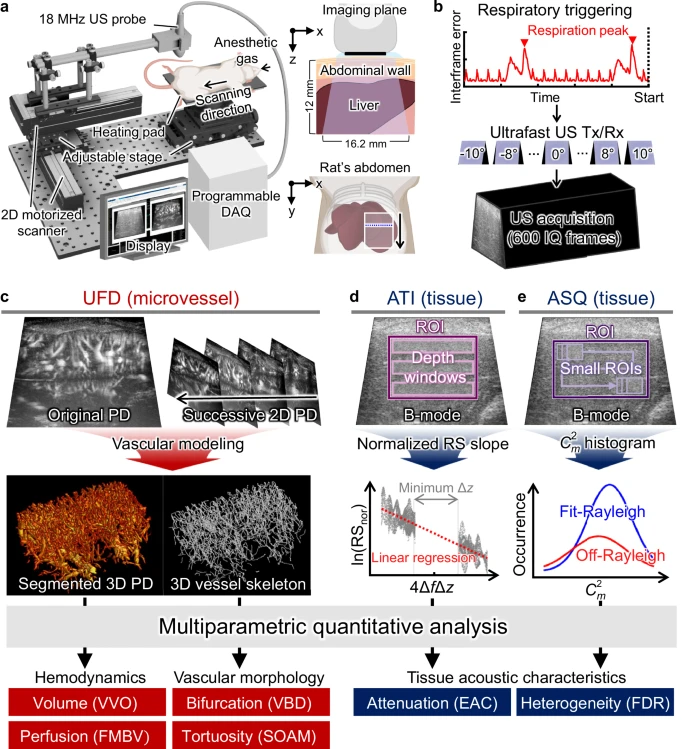
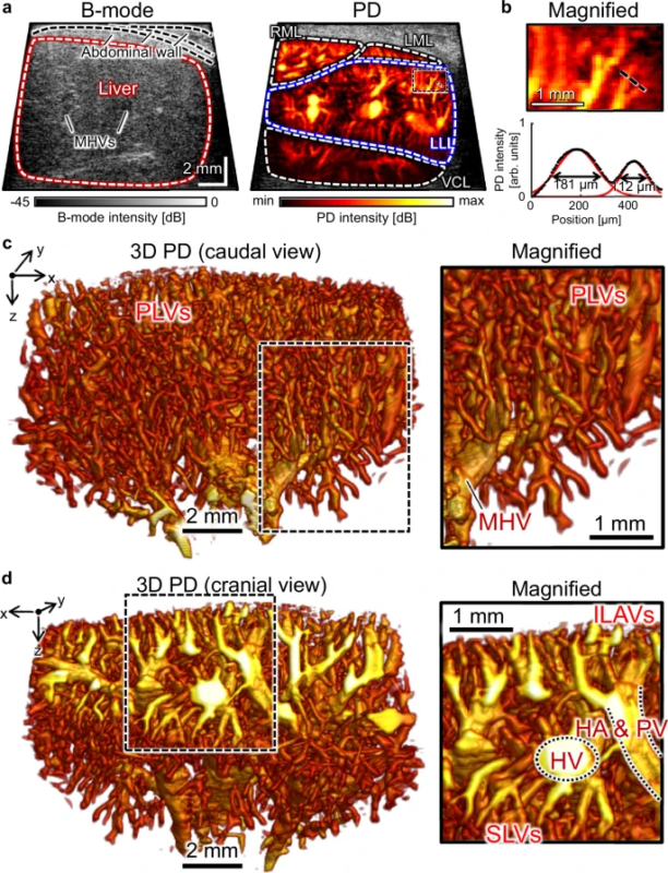
Fig. 2: In vivo 3D UFD image of hepatic vasculature (Highlights the intricate network of blood vessels)
How Hepatologists Use Liver Biopsy in the Evaluation of Liver Disease?
Hepatologists do a liver biopsy to get small samples from the liver. This helps them look closely at liver damage and see how liver disease is developing. A liver biopsy can help find out if someone has steatotic liver disease and check how bad fibrosis is. It is also useful for making choices about the right treatment. A liver biopsy is still an important step when doctors need a full picture of a person’s liver disease.
3D monitoring of SLD progression
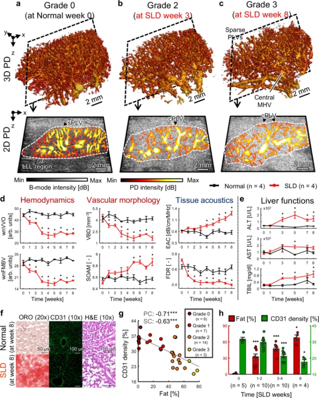
Shows changes in vascular indices over 8 weeks
The use of figures helps us better understand the changes that happen in the liver as steatotic liver disease gets worse. This approach looks at many things at once. It shows details about blood flow and other liver features. With this, healthcare pros get a full picture of steatotic liver disease and how it might respond to treatment.
Clinical implications and future potential
The study shows there is a strong link between checking blood vessels and how bad fat can build up in the liver. This problem is called fatty liver disease, or steatotic liver disease. The team used many ultrasound tests and some machine learning to help with this work. They made one main score from the ultrasound results. This score showed how serious liver disease is. It gave the right answer about 92% of the time when used on people with fatty liver or steatotic liver disease.
Professor Chulhong Kim said ultrasound with ultrafast Doppler shows more than regular scans. This way, doctors get to see little changes in blood vessels that they may not catch otherwise. It helps them know more about what is going on when they check a patient. Using ultrasound like this can help them find health problems sooner and bring better results.
Professor YongJoo Ahn said that if we can notice and use small changes in blood vessels early, it will help make medicine better and more exact. He also thinks that this can help people get care for other liver diseases in the future.
This study was done by a team led by Professor Chulhong Kim and Professor Yong-Joo Ahn at POSTECH. Professor Kim works in the Departments of Electrical Engineering, IT Convergence Engineering, Mechanical Engineering, and the Graduate School of Convergence Science and Technology. Professor Ahn is in the Department of IT Convergence Engineering and the Graduate School of Convergence Science and Technology.
Conflict of interest statement
The research in this blog has no competing interests. Professor Chulhong Kim does have financial connections to OPTICHO. However, he states the company did not take part in the work. All other authors also say they have no conflicts of interest for this study.
MeSH terms and classification
The main MeSH terms for this research are “Fatty Liver,” “Ultrasonography,” “Liver Diseases,” and “Magnetic Resonance (MR) Imaging, Three-Dimensional.” These groups help people sort and find this research in medical articles. They make it easy for other researchers and clinicians to read about new ways to spot steatotic liver disease. This is good for work on liver disease, fatty liver, and medical tests that use magnetic resonance or MR.
Grants and funding sources
This research got help from the National Research Foundation of Korea (NRF) with grants 2023R1A2C3004880 and RS-2024-00335346. The study shows how funding is key to moving ultrasound forward. It also helps add to what people know about checking liver diseases. The team used new ways of imaging, like MRI, ultrasound, and CT, to study the liver in vivo.
References and citation management
Managing your citations in the right way helps share your research and its findings. When you use the American Medical Association (AMA) style or other citation styles, it is important to be sure every source in this study is listed clearly. That way, future researchers can find the sources you used and add to the ideas you share in this blog. Good tools for citation are My Bibliography and PubMed. These tools help authors and researchers keep track of citations easily and make the work simple for everyone.
Source: Nature Communications

